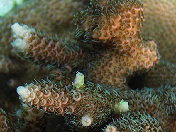
same coral one month later.
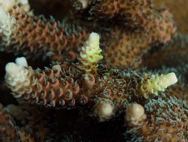
Picture taken under PC Actinic
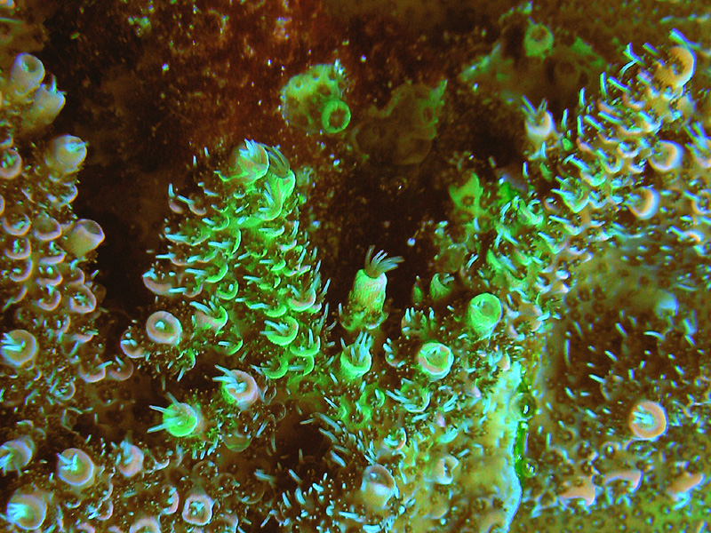
below is a close up detail of the spreading green pigment inside the corals tissue. Picture taken on cloudy day with 20K halide.
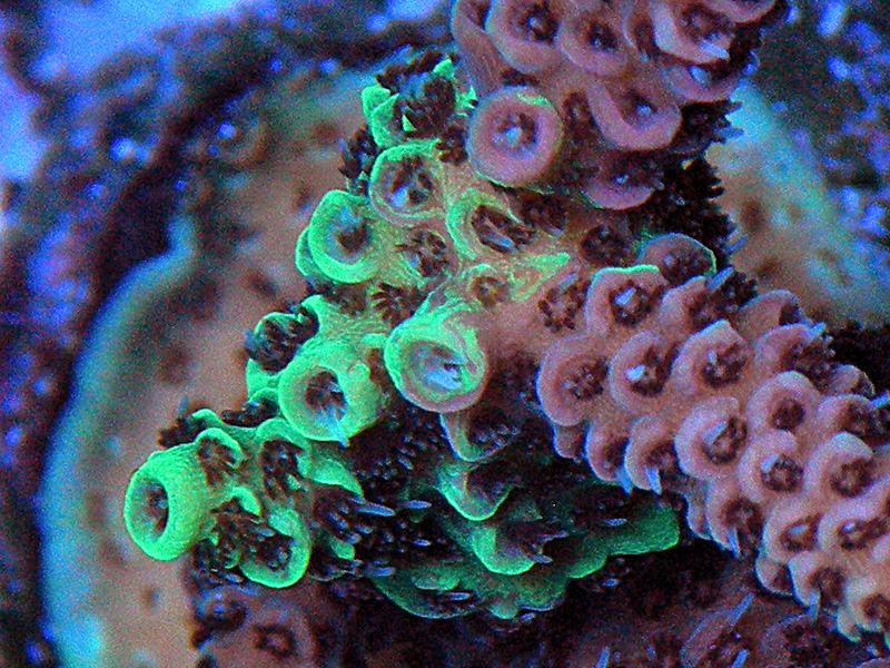
below is a shot of the underside of a pink millepora showing an area of green florescent pigment
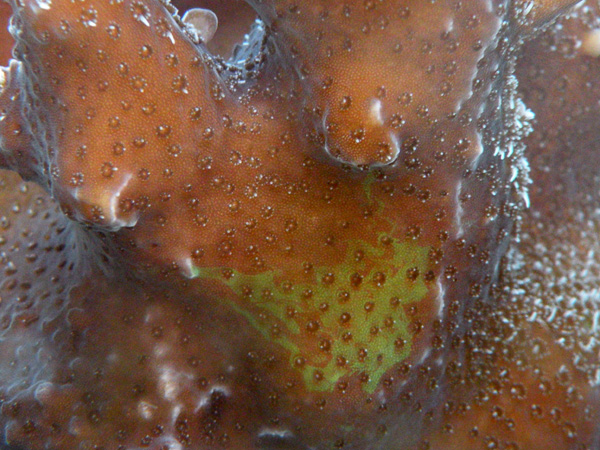
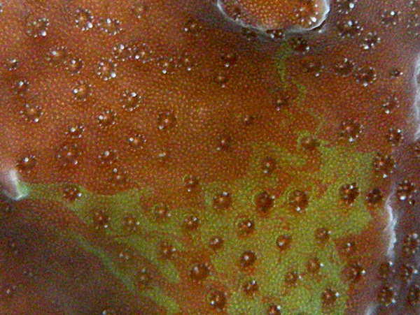
below is a close up showing the swirling pattern as the green pocillopora pigment slowly migrates through the coral
